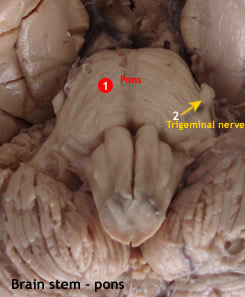
| The Pons |
|
 |
The pons consists of two parts i.e
- Ventral basal pons
- Dorsal pontine tegmentum
The basal pons makes a ventral bulge with a median basilar sulcus which lodges the basilar
artery.
The pons contains cranial nerve nuclei, ascending and
descending tacts, many pontine nuclei including an extensive reticular formation.
Cranial nerves attached close to the pons include:
- Trigeminal:
Junction of pons and middle cerebellar peduncle.
- Abducent: Junction
of pons and pyramid.
- Facial: Junction
of pons with olive.
- Vestibulocochlear:
Crebellopontine angle.
|
| |
|
 |
Sections through the pons
and pontine tegmentum characterized by the presence of the following:
- Dorsal fourth ventricle - dorsal.
- Longitudinal descending fibers - ventral.
- Transverse fibers to the cerebellum - ventral.
- Pontine nuclei - ventral.
- Reticular formation - dorsal.
- Medial lemniscus fibers - dorsal.
- Vestibular nuclei - dorsolateral.
- Spinothalamic fibers.
- Nuclei of trigeminal, abducent, facial nerve.
- Trapezoid body and nucleus.
- Locus ceruleus.
|
| |
 |
| |
| |
|
|
|
The cranial nerve nuclei include
- abducens
- Facial
- Motor part of trigeminal
- Chief sensory nucleus of trigeminal
- Vestibular nerve
Other nuclei include the following
- Pontine nuclei. These relay information from the neocortex to the cerebellum. Corticopontine fibres terminate in the pontine nuclei, and pontocerebellar fibres arise from these nuclei and enter the cerebellum as the middle cerebellar peduncle.
- Dorsal nucleus of trapezoid body (superior olivary). This acts as a relay station for some fibres of the cochlea projection. Some of the axons eventually synapse in trochlear an oculomotor nuclei.
- Pontine reticular formation nuclei. These control lacriminal glands (lacriminal nucleus) or salivary glands (superior salivatory) or control cardiorespiratory function.
Fibre tracts include the following
- Corticospinal continuing into the pyramid
- Corticonuclear terminating on CN, V, VI and VII.
- Corticopontine
- Trapezoid body which eventually forms lateral lemniscus
- Ascending tracts – spinothalamic, medial lemniscus
- Interconnecting fibres such as medial longitudinal fasciculus (MLF)
|
|
| Blood supply to pons |
This is mainly from pontine branches of the basilar artery.
These are grouped into 3:
i) Paramedian branches which supply the medial pontine region encompassing:
(+) Pontine nuclei
(+) Corticopontine
(+) Corticospinal
(+) Corticobulbar tracts
ii) Short circumferential arteries supplying adjacent anterolateral part of the pons and variable parts of the overlying tegmentum.
iii) Long circumferential arteries, which supply lateral parts of middle cerebellar peduncle, and most of the pontine tegmentum.
iv) Anterior inferior cerebellar artery supplying caudal pontine tegmentum. |
|
| Vascular lesion |
|
 |
Vascular lesions of the pons may result from occlusion of a pontine branch of the basialr artery.
The area of infarction may involve the following structures and hence the respective disorders:
a) Corticospinal fibres: contralateral hemiparesis.
b) Abducens nerve: Paralysis of ipsilateral rectus muscle with medial strabismus or squint. |
|
| Cerebello Pontine Angle |
|
| |
- Angle between the pons and the cerebellum.
- Bounded by:
(+)
Pons - rostrally
(+) Medulla - caudally
(+) Cerebellum -laterally
- Two nerves found in that interval i.e. the facial and vestibulocochlear.
Tumors at the CP angle compress CN VII and VIII, together with the pyramid and cerebellum. They present with CN VII/VIII defects together with motor deficits |
| |
| |